CLINICAL
Overview
Sampling techniques for standard microbiological analysis (i.e. wound culture) include swabbing the wound bed, needle aspiration and deep-tissue biopsy.[1][2] This topic provides step-by-step guidance on how to collect a superficial wound swab for wound culture and susceptibilities (C&S) when wound infection is suspected. For a review on when to collect a specimen for wound C&S and comparison of specimen collection methods (i.e. wound swab, tissue biopsy and needle aspiration), see topic "Wound Culture - Swabs, Biopsies, Needle Aspiration". For guidance on when and how to perform a wound biopsy see topic "How to Perform a Wound Biopsy".
Background
Definitions
- Wound culture: wound cultures are laboratory tests to determine the type and quantity of microorganisms in a wound. To perform a wound culture, a sample of tissue or fluid taken from the wound bed and placed in a sterile container for transportation to the laboratory. In the laboratory, the sample is placed in a substance that promotes growth of organisms and the type and quantity of organisms that grow are assessed by microscopy.[2]
- Susceptibility study: if upon wound culture pathogenic microorganisms are identified, a susceptibility study is performed at the laboratory by growing the pure bacterial isolate in the presence of varying concentrations of several antimicrobials and then examining the amount of growth to determine which antimicrobials at which concentrations inhibit the growth of the bacteria.[3]
- Wound swab for culture: wound swab is one of the sampling techniques used for wound culture. Other techniques include needle aspiration and tissue biopsy.[1][2] Although laboratories often prefer tissue or aspirate as specimens for microbiological wound culture (as these yield more microbes) [4], those specimens are not as easily obtained as wound swabs.[1] On the other hand, while wound swabs are minimally invasive, inexpensive, easier to perform, and widely utilized in clinical practice, they often fail to detect biofilm infection or pathogenic strains invading deeper tissues.[1] Wound swabs are collected using different techniques, such as the Levine technique and the Z technique [5][6]:
- The Levine technique involves rotating the wound swab over a 1cm2 area of the wound [6]
- The Z technique involves rotating the swab between the fingers in a zigzag fashion across the wound without touching the wound edge [6]
- Comparative studies show that the Levine technique is superior to the Z-swab technique for both acute and chronic wounds (P≤ 0.001 for both types of wounds).[2][5][6][7] Furthermore, the Levine technique when performed correctly may help decrease the chance of collecting skin contaminants that will interfere with culture results.
Relevance
- All chronic wounds are contaminated by several types of bacteria.[7][8] Patients with chronic wounds should undergo a comprehensive assessment.[2] If there are signs and symptoms of local infection, spreading infection or systemic infection and/or delayed healing, a wound culture is indicated to determine the microorganisms causing infection and to guide antibiotic therapy.[1][2] For signs and symptoms of local infection, see topic "Wound Culture - Swabs, Biopsies, Needle Aspiration".
- Among wound culture specimen collection techniques, wound swab is most frequently used due to its practicality and accessibility despite its limitations.[1][2]
- Prior to collecting a wound swab, wound cleansing without antimicrobials and wound debridement should be completed in order to remove debris and necrotic tissue from the wound bed.[2] Not doing so will increase the likelihood of collecting contaminants, which will interfere with culture results.
- When interpreting culture results, it is important to do so in light of clinical assessment and judgement. Wound swabs (even when collected with the Levine technique) have a sensitivity of 80-90% and a specificity of ~55%, that is, they may fail to detect or rule out wound infection. See section 'Wound Culture Techniques' in topic "Wound Cultures - Swabs, Biopsies, Needle Aspiration".
Care Setting
- Wound swabs may be performed in a multitude of care settings, including inpatient hospital, outpatient hospital, home health, skilled nursing facilities and others.
Scope of Practice for Collection of a Wound Swab
- Wound swab collection for culture is within the scope of several healthcare professionals including physicians, nurse practitioners, physician assistants, nurses, and physical therapists. In the United States (U.S.), medical assistants may be allowed to collect a wound swab for culture depending on each state's laws and regulations.[9]
- In the U.S., an order should be obtained for wound culture and susceptibility studies (C&S) from the treating physician/nonphysician practitioner (as required by CFR, Title 42, Volume 2, Chapter IV, Part 410.32(a) Ordering diagnostic tests).[10]
- If conservative sharp wound debridement is necessary to expose an area of viable tissue before collecting a wound swab, the procedure should be within the scope of the clinician performing the debridement. See scope of practice in topic "How to Perform Conservative Sharp Wound Debridement".
General Indications and Contraindications
Indications
According to the International Wound Infection Institute (IWII) consensus, collection of a wound specimen with a wound swab using the Levine technique is indicated for initial monitoring of wounds with suspected infection, including [2]:
- Acute wounds with classic signs and symptoms of infection
- Chronic wounds with signs of spreading or systemic infection
- Chronic wounds with signs of local wound infection and/or delayed healing
- Collection of specimen prior to institution of antibiotic therapy is preferred.
Other indications include [2]:
- To be in compliance with local protocols for the surveillance of drug-resistant microbial species
- Wounds where the presence of certain species would negate a surgical procedure (e.g. beta hemolytic streptococci in wounds prior to skin grafting)
For infected wounds that have failed to respond to antimicrobial intervention, or are deteriorating despite appropriate antimicrobial treatment, the IWII recommends obtaining a tissue biopsy for C&S. See topic "How to Perform a Wound Biopsy".
Contraindications
Contraindications to wound cultures in general include:
- "Routine" wound cultures. Wound cultures should not be undertaken routinely or without substantial cause (e.g. no signs of infection or delayed healing). Exceptions include the need to local protocols that require screening of drug-resistant organisms, or if the presence of certain species would negate a surgical procedure.[2]
- Inability to transport the culture within 4-24 hours of collecting the specimen.[2][11][12]
- Specimens collected from fresh bites, as infectious agents may not be recovered.[12]
- Wounds covered with slough, debris and necrotic tissue. Cleansing and debridement should be completed before specimen collection, so as to recover species at and below the wound surface.[2]
- Previous wound culture performed within the last 24-72 hours. The usual laboratory turnaround time for wound cultures can take up to a week (3-5 days for aerobic, 4-7 days for anaerobic culture)[11][13]
Considerations when deciding to collect a specimen with a wound swab:
- For patients with advanced stage pressure ulcers/injuries (stage 3 or 4), culture of a superficial ulcer swab might not be enough to accurately diagnose infection or guide antibiotic therapy. If feasible, it is preferable that culture be performed using specimens collected during surgical debridement (i.e. tissue biopsy).[14]
- Rationale: A study that compared culture results of superficial swabs and intraoperative specimens of 116 patients with advanced stage pressure ulcers/injuries (PUs/PIs) showed that the concordance between superficial swabs and intraoperative specimen culture was found only in 25 out of 116 cases (22%).[14] For those patients, prescribing antibiotics based on swab culture results may lead to unnecessary antibiotic administration in case of a false-positive result, or to insufficient antibiotic therapy in case of an unrecognized osteomyelitis.[14]
- If osteomyelitis is suspected, definitive diagnosis is achieved with a bone biopsy for culture and histology.[15][16] However, probable diagnosis through positive results of a combination of diagnostic tests (e.g. plain X-ray, magnetic resonance imaging, or radionuclide scanning) is acceptable, as bone biopsies are not always feasible or practical. For more on diagnostic tests for osteomyelitis see section 'Diagnosis: Diabetic Foot Infection - Osteomyelitis' in topic “DFU Intro and Assessment”.
HOW TO COLLECT A WOUND SWAB (LEVINE TECHNIQUE)
Assessment
- Prior to collecting a wound swab, it is important that a comprehensive patient and wound assessment be performed. See topic "How to Assess a Patient with Chronic Wounds" and "How to Determine Healability of a Chronic Wound".
Procedure: Wound Swab for Culture and Susceptibility (Levine Technique)
After determining that collection of a wound swab is indicated, clinicians may utilize the following steps as a guide for collecting a wound swab using the Levine technique.
Tables 1 below illustrate steps and rationale to appropriately collect a wound swab with the Levine technique.[2][17][18]
Table 1. Pre-procedure
| Steps | Rationale/ Tips |
1. Confirm presence of an active order for wound culture by a qualified healthcare provider
| - This ensures compliance in settings where individuals who are not qualified by licensure to order the procedure have active orders to perform the procedure.
|
2. Ensure that the wound swab can be taken to the laboratory within 24 hours
| |
3. Ensure correct patient identity per protocol.
| - Facility policy will dictate how patient should be appropriately identified.
|
4. Inform and seek permission from patient to obtain specimen or confirm presence of informed consent
| - By obtaining informed consent, the patient is aware of the purpose for the procedure, benefits, as well as the expected outcomes and potential complications.
|
5. Ensure adequate assistance, supplies, and equipment:
- 2 sets of clean gloves (for cleansing the wound and taking the swab)
- 1 set of clean or sterile gloves (to apply the new dressing depending on the technique used)
- 100 ml sterile normal saline or sterile water plus equipment and supplies needed to cleanse the wound
- Sterile swab kit for C&S (culture for aerobic bacteria only).
- If there are 2 or more wounds in the same location use a separate swab kit for each wound.
- Biohazard transport bag and laboratory requisition
- Appropriate supplies to redress the wound
| - A sterile or no-touch aseptic technique is used when taking a wound swab for C&S
|
6. Ensure patient is positioned for comfort. Assess for pain and pre-medicate if necessary prior to the procedure.
| - Ensure the wound to be sampled is easily accessible, and that assistance to position the patient (if needed) is available prior to starting the procedure
|
Table 2. Procedure
| Steps | Rationale/Tips |
1. Perform hand hygiene, and don clean gloves
| - A sterile or no-touch aseptic technique is used when taking a wound swab for C&S
|
2. Open supplies onto separate surface or tray, in an area independent of patient or clinician movement.
| - Tools should never be placed on an unstable surface, such as the patient; this could potentially cause injury.
|
3. Remove the soiled wound dressing, if present.
|
|
4. Remove the gloves and perform hand hygiene. Put on clean gloves.
|
|
5. Cleanse the wound with normal saline (do not use antiseptics), pat dry with sterile gauze
| - It is important to recover species at and below the wound surface, therefore wound cleansing should be completed before sampling the wound
|
6. If excessive debris and/or necrotic tissue are present, remove them through conservative sharp debridement (if knowledge, skills and license permit). See topic "How to Perform Conservative Sharp Wound Debridement" 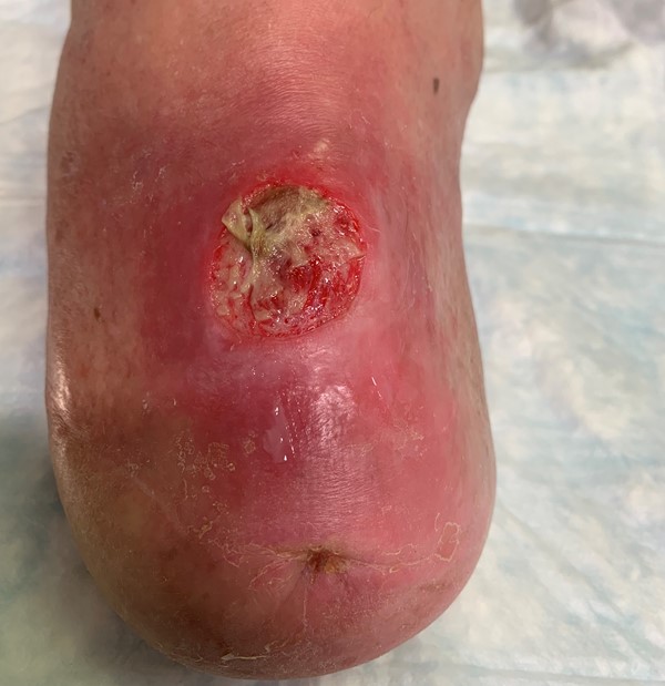
Fig. 1. Ulcer pre-debridement 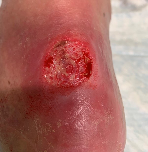 Fig. 2. Ulcer post-debridement | - It is important to recover species at and below the wound surface, therefore debridement (if necessary) should be completed before sampling the wound
|
7. If conservative debridement is performed, cleanse wound bed post debridement with saline and pat dry with sterile gauze
|
|
8. Identify the healthiest, cleanest looking area of granulation tissue. Do not choose areas with exudate, pus, devitalized tissue, tunneling or undermining
| - Minimize collection of contaminants, as those will interfere with culture results
|
9. On the identified area, rotate the end of the sterile applicator (swab) over a 1cm2 area. For at least 5 seconds, apply enough pressure to express fluid from within the wound tissue. If you cannot identify a 1cm2 area, do not take the specimen and notify the provider 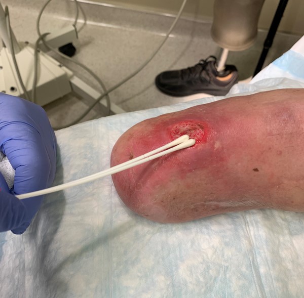
Fig. 3. Collecting a wound swab | - If the wound is relatively dry, collect the specimen with two cotton-tipped swabs moistened with sterile non-bacteriostatic saline
|
| 10. Insert swab into sterile tube with transport medium and send to lab immediately | - The swab should be placed in a bacterial swab transport (e.g. ESwab, Amies Agar gel transport media)[19]
- Some swab transports may have ESwab. ESwab is a liquid-based, multipurpose open platform, collection and preservation system that maintains viability of aerobic, anaerobic and fastidious bacteria for up to 48 hours at refrigerator and room temperature.[19]
- Ensure the swab tip is in contact with the transport medium at the base of the tub
|
| 11. Remove gloves. Put on clean or sterile gloves (depending on the wound dressing technique) | - Moisture management and wound bed preparation principles should be adhered to
|
| 12. Apply wound dressing and remove gloves. |
|
Table 3. Post-procedure
| Steps | Rationale/Tips |
Prepare the specimen for laboratory submission. Include the following information in the laboratory requisition and/or specimen label [2]: - Time of collection
- Information on the wound: anatomical location, duration and etiology, unusual organisms (e.g. Nocardia) if suspected
- Information on the patient: demographics and relevant comorbidities
- Clinical indication for the wound sample: signs and symptoms, and suspected microbes
- Current or recent antibiotic use
| - If more than one swab is collected from two or more wounds in the same location, specify the wound by documenting the location or another identifier on the specimen container.
- Samples that are usually not accepted for culture by laboratories: syringes with needle attached, dry swabs, refrigerated or frozen samples
|
- Place the specimen in a biohazard transport bag and transport to the lab as soon as possible.
- If the swab cannot be sent to the lab within 24 hours discard it and collect a new C&S swab.
| |
Documentation
Documentation should include:
- Wound assessment and characteristics (e.g. appearance, exudate, signs of infection)
- Medical record documentation supporting medical necessity (i.e., for Medicare beneficiaries, CMS requires use of at least one of the ICD-10-CM code(s) that support medical necessity. A sample list is provided by the Local Coverage Article: Billing and Coding: Susceptibility Studies (A57176)).[10]
- A wound C&S order from the treating physician/nonphysician practitioner (as required by CFR, Title 42, Volume 2, Chapter IV, Part 410.32(a) Ordering diagnostic tests).[10]
- Description of the specimen collection procedure, patient's tolerance to the procedure and response to analgesia (when used).
- Labeling of the specimen container and completion of the laboratory requisition with the following information:
- Time of collection
- Information on the wound: anatomical location, duration and etiology, unusual organisms (e.g. Nocardia) if suspected
- Information on the patient: demographics and relevant comorbidities
- Clinical indication for the wound sample: signs and symptoms, and suspected microbes
- Current or recent antibiotic use
Video: How to Collect a Wound Swab (Levine Technique) for Culture
Video 1. Levine swab culture technique. Patient with diabetes and chronic ulcer on the left elbow (By Scott Robinson, MD)
HOW TO ASSESS EFFECTIVENESS
Test limitations
- Even when adequately collected with the Levine technique, wound culture of superficial wound swabs may not be effective in detecting or ruling out infection. Wound culture from wound swabs have a sensitivity of 80-90% and a specificity of ~55%.[14][20] See section 'Wound Culture Techniques' in topic "Wound Cultures - Swabs, Biopsies, Needle Aspiration". As a result, clinicians should be wary of interpreting wound culture results/report in isolation. Consider results/reports in the context of the individual, their wound and your clinical judgement. If appropriate, consult a microbiologist or an infectious disease expert.[2]
- Wound swabs often fail to detect biofilm infection or pathogenic strains invading deeper tissues.[1][2]
- Chronic wounds are usually initially tested for aerobic bacteria. Testing for fungi and anaerobic bacteria requires additional specimens, investigations and processing.
- Because standard techniques have limitations that prevent many microorganisms from being cultured, new molecular and imaging technologies have been developed but those are not yet widely available (e.g. PCR assays, DNA sequencing techniques).[2] See section 'Limitations' in topic "Wound Cultures - Swabs, Biopsies, Needle Aspiration".
Management after wound swab collection
- After collection of specimen for culture, depending on the patients condition, patients can be started on empiric antibiotic therapy or followed up within 24 hours, or followed up within 3-5 days and then be started on antibiotics based on culture results if clinically indicated. Close monitoring of the patient and wound is important. For instance, if the patient is able to reliably report any complication or worsening of symptoms over a televisit or phone call, an in person follow up appointment may be scheduled in 1 week. If patient has multiple comorbidities or arterial insufficiency, follow up within 24-48 hour may be appropriate. Table 4. illustrates management of a patient with suspected wound infection, after collection of a wound swab.
- Interpreting wound culture results/microbiology report: the diagnosis of infection is primarily clinical and wound culture is a test to confirm clinical diagnosis.
- Swab results are usually reported as the number of organisms per swab.[1]
- If pathogens are detected and sensitivities are provided, the IIWC recommends clinicians consider clinical indications before starting antibiotics.[2]
- If an infected wound does not respond to appropriate treatment with culture-guided antibiotics, consider additional factors that may impact wound healing before re-culturing a wound.[2]
Table 4. Management of a patient with suspected wound infection, after wound swab collection
Management of the patient depicted in Video 1 above, after wound swab collection:
- Clinical presentation: Video 1 above shows a patient with diabetes, smoker and a chronic ulcer located on the left elbow, near the ulnar nerve. Clinical signs of infection included erythema in the periwound area and a tingling sensation along the distribution of the ulnar nerve, down to the 5th finger.
- Wound swab and empiric antibiotics: after the wound was cultured, the patient initiated a course of empiric antibiotic therapy with Trimethoprim / Sulfamethoxazole, and asked to return for a follow up appointment in a week.
- Wound culture results: even though the Levine swab culture technique was followed, the culture of the specimen did not grow any microbes.
- Follow up appointment: upon follow up the erythema and tingling sensation had subsided after implementation of standard wound care, topic antimicrobials and empiric antibiotic therapy. If symptoms had worsened, a punch biopsy for quantitative analysis would have been indicated.
In summary, it is important to note that sometimes swab culture results might be negative despite clinical signs of infection. A negative culture result in this case emphasizes the importance of taking the clinical picture into consideration. |
CODING, COVERAGE, REIMBURSEMENT
- There is no CPT code for the swab of a wound, it is included in the evaluation and management (E/M) charge. Adequate documentation of the procedure will help justify a higher level of E/M. See topic "2021 Office/Outpatient E/M Services Updates for Wound Care and HBOT".
- If conservative sharp wound debridement is performed before collecting a wound swab, this procedure may be billed instead of the E/M. See topic "Coding and Billing Essentials in Wound Care".