INTRODUCTION
Overview
Wound care clinicians often face challenging ulcers that have been previously treated with little or no success by other healthcare providers. Some of these ulcers are related to cutaneous vasculitis. This topic describes how an atypical lower extremity ulcer was diagnosed as new-onset cutaneous vasculitis at a wound care clinic. For an updated, summarized review on assessment and management of this often misdiagnosed condition, see topic "Cutaneous Vasculitis". For a list of other types of inflammatory ulcers, see topic "Inflammatory Ulcers".
CASE
Background
A 28 year old white male, truck driver, presented to the wound clinic with a necrotic ulcer in the left leg for 2 weeks. The patient had a history of diffuse pain and swelling in both legs for 3 months, which intensified 2 month after onset, prompting the patient to visit the Emergency Department (ED). Upon examination, he had a tender, erythematous cutaneous lesion of 0.3 cm in diameter in the anterolateral aspect of the tibia in the middle third of the left leg. No fluctuation areas that suggested abscess were immediately visible. Both calves were painful upon palpation, and dorsiflexion of the foot (positive Homan’s sign). 1+ pitting edema was found bilaterally. The patient was afebrile and had no other systemic manifestations. To rule out a possible deep venous thrombosis (DVT), a soft tissue ultrasound of both legs was performed, and while no signs of DVT were found, a 1 cm pocket of fluid under the erythematous area was detected. Suspecting of abscess, the ED provider incised the skin under local anesthesia and drained the fluid, which came clear. Bacteriological culture of the fluid showed no growth of any pathogenic organism. Patient was prescribed oral cephalexin and referred to the wound clinic.
The patient had a history of seasonal allergies, took occasional oral cetirizine and was otherwise healthy (no history of malignancy, recreational drugs or infection).
Physical examination and workup
At the first visit to the wound clinic, patient presented with a necrotic lesion to the anterolateral aspect of the left leg, which expanded from the previously linear drainage incision site to a 3 x 2 cm necrotic crust, surrounded by erythematous tissue and non-blanchable, purpuric lesions over a 4 cm radius around the necrotic area (Figure 1). On the right leg, the patient had been wearing a CAM walker prescribed by an orthopedist due to a “pulled muscle” for 2 weeks.
At this first visit, necrotic tissue of the ulcer was removed (Figure 2) and the ulcer was covered with and antibacterial collagen dressing. At the follow up visit, the lesion seemed to be larger.
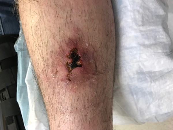
Fig. 1. Necrotic lesion on the anterolateral aspect of the left leg
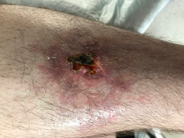
Fig. 2. Necrotic tissue of the ulcer being removed
Diagnosis and differential diagnosis
At the follow up visit, cutaneous vasculitis was considered as a potential diagnosis and a biopsy of the perinecrotic area was taken. Laboratory tests were ordered with results as follows:
- Complete blood count (CBC): normal range
- Rapid plasma reagin (RPR): non reactive
- Rheumatoid Factor (RF): normal range
- Anti-nuclear Antibody (ANA): normal range
- Erythrocyte Sedimentation Rate (ESR): elevated
- C Reactive Protein (CRP): elevated
- Liver and renal function: normal range
Treatment and follow-up
Patient was empirically prescribed a course of oral prednisone dose (20 mg two times a day for 1 week), followed by tapered doses. Pathology report confirmed presence of leukocytoclastic vasculitis (LCV, a term that defines vasculitis of the small vessels in which the inflammatory infiltrate is composed of neutrophils).
A week later, patient showed improvement with oral prednisone and was referred to a rheumatologist for further workup and refinement of diagnosis among possible causes of cutaneous vasculitis.
DISCUSSION
Wound care specialists often face challenging ulcers that have been previously treated with little or no success by other healthcare providers. Some of these ulcers are related to cutaneous vasculitis. One of the most frequent challenge in practice is to clinically suspect, diagnose, and identify whether the vasculitic ulcer secondarily results from a probable etiology (e.g. drugs, infections, sepsis, autoimmune diseases, etc), or from impairment of the cutaneous microcirculation due to primary vasculitic inflammation.
Clinical presentation
Cutaneous vasculitis can be multifactorial and present with a variety of clinical findings. These causes need to be addressed in order to promote ulcer healing. Investigation for underlying conditions such as infections, medications, connective tissue diseases, and malignancy should be considered, as these account for 23, 20, 12, and 4 percent of cases of cutaneous vasculitis, respectively.[1] Primary systemic vasculitides account for only 4 percent of cases. Even after further investigation, the cause of vasculitis is unidentifiable in 3 to 72 percent of patients.[1] See section 'Assessment' in "Cutaneous Vasculitis"
Diagnosis and differential diagnosis
Vasculitis should not be overlooked as a consideration in the differential diagnosis of new-onset lower extremity ulcers. Among other conditions, ulcers resulting from vasculitis can resemble venous leg ulcers (VLU), fungal infections (e.g. sporotrichosis), atypical trauma (e.g. puncture wounds from contaminated sources, splinters, locust thorns, etc), calciphylaxis ulcers, or ulcers caused by spider bites/other envenomations. VLUs commonly presents in between the mid-calf and approximately 1 inch below the malleolus. The recurrence of an ulcer in the same area is highly suggestive of venous ulcer. History of puncture may be helpful in atypical trauma or fungal (rose thorn in sporotrichosis). History of spider bite may be present but often this is unknown or uncertain, but skin findings are usually temporally related, usually ulcers develop within hours to a couple of weeks after the bite or envenomation. Because other diseases may have similar presentations, histopathologic examination is necessary to confirm diagnosis.
For the diagnosis of cutaneous vasculitis, a recent lesion (24-48 h old) is appropriate for routine biopsy with H&E. The biopsies for direct immunofluorescence should be taken from the lesion, between 8 and 24 h of age. Immunofluorescence analysis for demonstration of immunoreactants in dermal vessels ideally requires biopsy of an early or histamine-induced lesion, because immunoglobulins may disappear over time.[2] Infections, insect bites, pyoderma gangrenosum, and even in secondary vascular changes underneath the ulcers may exhibit leukocytoclastic vasculitis; therefore, a clinicopathological correlation is necessary before establishing a final diagnosis. The possibility of systemic involvement should be thoroughly searched.[3] Furthermore, an infection, medication, or a systemic disease such as systemic lupus erythematosus must be searched out before reaching a definitive diagnosis.[3] See more information on biopsies and laboratory tests in the section 'Diagnosis' in "Cutaneous Vasculitis"
Treatment
Once systemic involvement has been excluded, the treatment of cutaneous small vessel vasculitis should be focused on symptom management. The majority of cases affect only the skin, are acute, and self-limited so aggressive immunosuppressive therapy is generally not advisable.
Identifiable triggers should be eliminated or treated. Prolonged standing should be avoided. Rest and elevation and use of compression stockings can be helpful to decrease stasis-related immune complex deposition and accelerate healing of ulcers on the lower legs. Analgesics should be used for pain control. Topical steroids can relieve itch or burning but do not prevent new lesions. More than half of patients require no systemic treatment.[4]
In complicated cases, like the one described here, systemic therapies are indicated. Any initial episode that is not self-limited and lasts longer than a few weeks likely warrants systemic treatment, even if it is relatively asymptomatic. Systemic corticosteroids, with initial doses of 0.5 to 1 mg/kg per day of prednisone, are recommended for those with severe, necrotic lesions or serious systemic manifestations, such as renal or gastrointestinal involvement. The response to such therapy is usually rapid, and the dose should be tapered slowly to prevent rebound. Long-term therapy may not be required if the process is self-limited. For refractory cases, treatment options include colchicine (0.6 mg 2–3 times per day, if tolerated) and dapsone (50–200 mg/d).[4] See more details on management of cutaneous vasculitis in the section 'Address patients' concerns' in "Cutaneous Vasculitis".
Key Takeaways
Some of the main takeaways from this case study are:
- One of the most frequent challenge in practice is to clinically suspect, diagnose, and identify whether the vasculitic ulcer secondarily results from a probable etiology (e.g. drugs, infections, sepsis, autoimmune diseases, etc), or from impairment of the cutaneous microcirculation due to primary vasculitic inflammation. Wound care clinicians need to have a high level of suspicion for cutaneous vasculitis when patients present with ulcers in the lower extremity but do not demonstrate an underlying anatomical/ functional alteration of large vessels.
- For cases of potential cutaneous vasculitis, it is important to rule out probable causes of the vasculitides (e.g, drugs, infections, autoimmune diseases, etc), even if the biopsy demonstrates leukocytoclastic vasculitis. Referral to a rheumatologist or vasculitis specialist for further workup and management is recommended.
|
Official reprint from WoundReference® woundreference.com ©2026 Wound Reference, Inc. All Rights Reserved
NOTE: This is a controlled document. This document is not a substitute for proper training, experience, and exercising of professional judgment. While every effort has been made to ensure the accuracy of the contents, neither the authors nor the Wound Reference, Inc. give any guarantee as to the accuracy of the information contained in them nor accept any liability, with respect to loss, damage, injury or expense arising from any such errors or omissions in the contents of the work.