CLINICAL
Overview
This topic provides a practical perspective on differentiating Stage 2 pressure ulcers/injuries on the gluteal region from selected common conditions. For a list of guidelines and quality measures related to PU/PI, see topic "Pressure Ulcers/Injuries - Overview". For an introduction and assessment of PU/PI including epidemiology, risk factors, etiology, pathophysiology, history, physical examination, diagnosis, differential diagnoses, documentation and ICD-10 coding, see "Pressure Ulcers/Injuries - Introduction and Assessment". For management of PU/PI, see topic "Pressure Ulcers/Injuries - Treatment". For a systematic approach to identifying patients at risk for developing pressure ulcers/injuries (PUs/PIs) and developing specific care plans, see topic "Pressure Ulcers/Injuries - Prevention". For best practices in care coordination, see topic "Pressure Ulcers/Injuries -Coordination of Care".
Background
- When individuals have skin breakdown on their gluteal region, it is often identified as a Stage 2 pressure ulcer/injury (PU/PI). However, not all skin breakdown in this area is related to increased pressure on the skin. Other common conditions include:
- Incontinence-Associated Dermatitis (IAD)
- Friction-Induced Skin Injury (FISI)
- Medical adhesive related skin injury (MARSI)
- Traumatic wounds (skin tears, burns, abrasions)
A few of the most common conditions are summarized and compared in the paragraphs below and in Table 1:
- Pressure Ulcers/Injuries: A pressure ulcer (PU), also known as pressure injury (PI), pressure sore, decubitus ulcer or bed sore, is an area of localized injury to the skin and/or underlying tissue, usually over a bony prominence or related to a medical or other device. A pressure ulcer/injury (PU/PI) can present as intact skin and/or ulcer and may be painful. It occurs as a result of pressure, or pressure in combination with shear.[1]
- Stage 2 PU/PI: According to the National Pressure Ulcer Advisory Panel (NPUAP) a stage 2 PU/PI is defined as follows: "Partial-thickness loss of skin with exposed dermis. The wound bed is viable, pink or red, moist, and may also present as an intact or ruptured serum-filled blister. Adipose (fat) is not visible and deeper tissues are not visible. Granulation tissue, slough and eschar are not present. These injuries commonly result from adverse microclimate and shear in the skin over the pelvis and shear in the heel. This stage should not be used to describe moisture associated skin damage (MASD) including incontinence associated dermatitis (IAD), intertriginous dermatitis (ITD), medical adhesive related skin injury (MARSI), or traumatic wounds (skin tears, burns, abrasions)"(Figure 1).[1]
- Incontinence-Associated Dermatitis: Incontinence-associated dermatitis (IAD) is a “form of irritant dermatitis that develops from chronic exposure to urine or liquid stool” (Figure 2).[2][3] IAD is one of four types of moisture-associated skin damage (MASD), which has has been defined as “inflammation and erosion of the skin caused by prolonged exposure to various sources of moisture, including urine or stool, perspiration, wound exudate, mucus, or saliva.”[2][3]
- Friction-Induced Skin Injury (FISI): A friction-induced skin injury (FISI) is an injury caused solely by friction, and not pressure.[4] Friction is defined as "rubbing of one body against another or the force that resists relative motion between two bodies in contact and/or material elements sliding against each other". One reason for the deletion of the words “and/or friction” from causes of PUs/PIs in the PU/PI definition by the NPUAP is to reinforce that skin injuries caused by friction are not to be considered PUs/PIs (Figure 3).[5] Friction in and of itself only causes superficial skin damage.[6]
- Friction injuries may be caused by acute or chronic abrasive/friction forces during sliding, scooting, or slouching behaviors. These behaviors are usually seen in individuals with impaired mobility, particularly when transferring and repositioning.[7]
Table 1. Comparison of stage 2 pressure ulcer/injury, incontinence-associated dermatitis and friction-induced skin injury [6]
| Conditions | Stage 2 pressure ulcer/injury
| Incontinence-associated dermatitis
| Friction-induced skin injury
|
| Pictures | 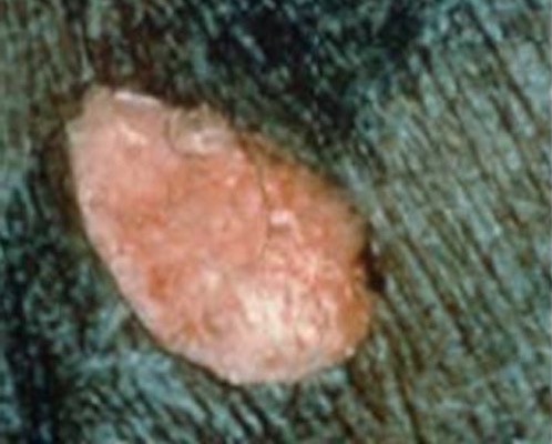
Fig. 1. Stage 2 PU/PI | 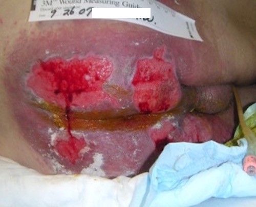
Fig. 2. Incontinence-associated dermatitis | 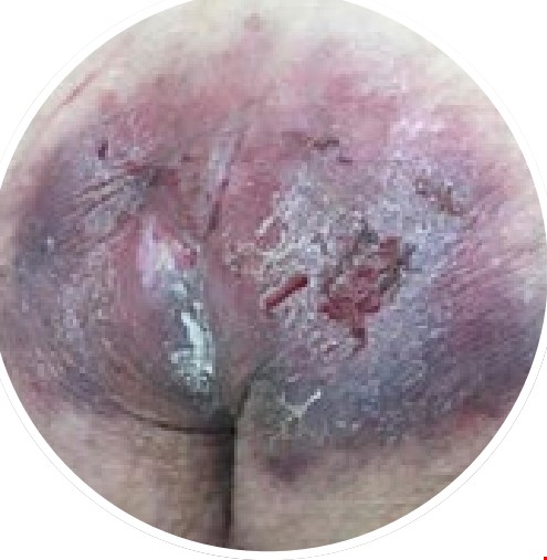
Fig. 3. Friction-induced skin injury[8] |
| Etiology | Pressure, or pressure in combination with shear | Caused by exposure of the skin to urine and/or feces
| Caused by friction, that is “the rubbing of two objects against each other when one or more are moving”
|
| Commonly affected areas | Usually over a bony prominence or related to a medical or other deviceA supine position causes coccyx/sacral PUs/PIsA sitting position causes ischial PUs/PIsPUs/PIs do not usually occur on the fleshy part of the buttocks as there is no bony prominence therePUs/PIs CAN occur on parts of the body that do not have a bony prominence due to an object (e.g. medical device). However, in order for lesion to be considered a PU/PI, clinicians must be able to identify the object causing the injury
| Most often found in the perianal area, buttocks, inner thighs and perineum
| Usually found over the fleshy/rounded part of the buttocks and posterior thighs |
Common presentations
| Shape is usually round or oval Depth: shallow lesions with red/pink wound bed without necrotic tissue. Not full-thicknessEdges: well-demarcatedShiny, pink or red open wound bed, no slough in wound bed. Can also present as serum-filled blistersPeriwound: non-blanchable erythema of intact skin | Shape is blotchy, not uniform, irregular
Depth: none or shallow, not full-thicknessEdges: irregular, diffuse, very shallowShiny, redness and skin irritation, no slough in wound bed. May also present with bleeding and be painfulPeriwound: may appear lighter or darker than surrounding skin, may be erythematous, erythema usually blanchable | Shape is irregularMultiple or single lesions
Depth: none, partial or full-thickness skin lossEdges: irregular. Chronic injuries have palpable changes including ridging, hyperkeratosis and hypertrophy of wound edgesWound is often dry. May be deep red if deeper injury or may be a shallow ulcer involving subcutaneous tissue. Not a deep crater, but may cover a large surface area. Some areas may have all the skin rubbed off (denuded) and appear open
Periwound: blanchable erythema of intact skin or chronic discoloration, hyperchromia, irritated, inflammed, edematous, lichenification, skin scaling. May bleed easily. |
| Key findings for diagnosis | - If the lesion is on a bony prominence, can the cause be linked to increased pressure, or pressure in combination with shear?
- If lesion is not on a bony prominence, the key is to be able to identify what caused the PU/PI (e.g., patient lying on a pulse oximeter connecter for hours). If cause can be linked to pressure, or pressure in combination with shear, lesion can be labeled as a PU/PI
| Redness or erythema of IAD is blanchable | - History of friction - friction to the buttocks can occur many times a day, for instance:
- Sliding a patient up in bed
- Sliding/scooting to the side of the bed to get up
- Scooting to the edge of a chair
- If caused by chronic friction, it presents with blanchable erythema or purple skin discoloration
|
| ICD-10 | - L89.x2 or L89.xx2, where "x" varies according to body location
- See all ICD-10 codes in section 'ICD-10' on "Pressure Ulcers/Injuries - Introduction and Assessment"
| - ICD-10 contains coding for diaper dermatitis, but does not contain a separate coding for IAD [9]
- L30.9 “dermatitis, unspecified" may be used for IAD [10]
| - T14.8xxA: Other injury of unspecified body region, initial encounter
|
Determining the etiology of the skin breakdown
Question the patient, family or caregivers regarding factors that may have led to the lesion:
- If bedbound, how often is repositioning done?
- Does the patient slide down in the bed? Scoot to the edge of the bed or chair?
- Is incontinence an issue?
Sometimes the cause is clear and your documentation will be easy. Sometimes it can be a combination of pressure, friction and/or incontinence. In those cases, clinicians should make sure documentation includes all causes of the patient's skin breakdown.
REVISION UPDATES
| Date | Comments
|
| 6/28/22 | Updated section on friction-induced skin injuries |