WoundReference improves clinical decisions
 Choose the role that best describes you
WoundReference logo

How to Apply Cellular and/or Tissue Based Products

How to Apply Cellular and/or Tissue Based Products

How to Apply Cellular and/or Tissue Based Products

ABSTRACT

This topic provides practical guidance with step-by-step pictures, protocol and tips on how to apply different types of cellular and/or tissue products (CTPs), also known as skin substitutes. For a comprehensive overview on cellular and tissue products from the clinical, coverage and reimbursement perspective, see "Cellular and/or Tissue Products". For guidance on selection of CPT see "How to Select Cellular and/or Tissue Products". For decision support on different CTP brands, see interactive feature matrices  “Human Skin Allografts”“Allogeneic Matrix”“Composite Matrix” and “Acellular Matrix”. For a Medicare-focused documentation checklist when using CTPs, see "Request for Cellular and/or Tissue Product"

   Videos

HOW TO APPLY

Steps and Rationale

The table below illustrates general guidelines for application of cellular and/or tissue products (CTPs) on wounds. Please refer to manufacturers' literature for specific instructions.  State-by-State Regulations on Conservative Sharp Debridement by Nurses in the United States How to Perform Conservative Sharp Wound Debridement

Process Tasks Rationale/ Tips
Prepare patient
1. Ensure wound is optimally debrided, with no clinical signs of infection and pre-treated with appropriate dressings. 
  • Assuming wound is refractory to standard care, consider aggressive debridement in conjunction with topical antimicrobial 2-3 weeks before CTP application as adjunct to wound bed preparation
  • Application of many CTPs is contraindicated on clinically infected wounds
  • For Medicare patients, review the requirements checklist "Request for Cellular and/or Tissue Product"

2. If needed, ensure patient is adequately medicated for pain prior to CTP application or change
  • For removal of previous dressings, please consider hydration of the dressing with saline, wound cleanser, or topical anesthetic agent to reduce patient discomfort during dressing change
Prepare for procedure
3. Please review safety information and instructional manual for the specific CTP before application. Do not apply product if patient has any contraindication
  • Be aware of your resources, set up an inservice from the product representative and keep their contact information handy.
  • Be familiar with the product indications, contraindications, and compatible therapies

4. Confirm there is an order for CTP and dressing change frequency
  • Having this information beforehand can avoid confusion 
Prepare wound
5. Remove previous dressing if applicable, ensuring all pieces are removed. Dispose per facility policy.
  • If a CTP has been previously applied, may gently remove non-adherent parts, but do not debride adherent parts

6. Non-viable tissue should be debrided as needed (See Video 1 below)
  • Wound healing will take place more rapidly if the wound is free of debris and devitalized tissue; mechanical debridement can be performed as needed during dressing changes to remove sessile tissue, but conservative sharp wound debridement may need to be performed for more adherent slough or eschar
  • See Video 1 below, see topic "How to Perform Conservative Sharp Wound Debridement"

7. Cleanse wound and periwound per orders or facility protocol. 
  • Ensure cleansing agent utilized is proven non-cytotoxic to healthy tissue

8. Assess wound for:
  1. Adequate hemostasis
  2. Exposed structures or sharp bony edges - some CTPs should not be applied on tendon, bones, nerves  (check feature matrix)
  3. Unexplored tunneling - some forms of CTPs may be applied in tunnels (e.g, flowable)
  • Attention to assessment of these factors will prevent adverse effects such as uncontrolled bleeding, tissue injury related to sharp bony edges, or untoward effects such as fistulization or injury of other structures in unexplored tunnels
Apply CTP
9. Using sterile technique, apply CTP as recommended by manufacturer
  • Different CTP presentations have different application instructions (e.g, powders, sheets, flowable). Follow manufacturer's instructions for use.
  • Remove CTP from packaging. The outer package is not sterile. The inner package is usually sterile (check manufacturer's instructions) and can be placed onto the sterile field.

10. Note CTP wastage for documentation/billing purposes
  • Medicare and many private insurers require documentation of how much product from a single package was used and not used during a single application
  • For documentation requirements, refer to the checklist 'Request for Cellular and/or Tissue Products' in topic "Cellular and/or Tissue Based Products"

11. Record the CTP lot number in the patient chart
  • For tracking of the medical device in case there is a recall or side effect
Secure the product
11. Secure CTP.  May use silicone contact layers, skin closure strips, petrolatum gauze, secondary dressings, negative pressure wound therapy, or compression bandage if treating venous ulcer (See Video 2 below)
  • CTP needs to be in close contact with the wound bed, with no air pockets so that it can act as a matrix and/or provide growth factors
  • See Video 2 below
Communicate care expectations
12. Communicate post-application instructions to patient, family and hand-off staff (home health, long term acute facility, skilled nursing facility, outpatient staff, peripheral providers)
  • Leave written instructions for next provider (or call), let patient and caregivers know how to manage post applicationadd link to post application instruction
Follow-up
13. Depends on the condition being treated and the type of CTP but in most cases, follow-up is scheduled in a week. 
  • Follow-up should be scheduled according to patient assessment considerations, clinical setting considerations, staff caring for patient, supplies available, and transportation reliability. 
  • TeleVisit Tool 2.0  for real time video/audio communication may be helpful in between visits
  • A circle drawn by provider on cover dressing can be used as visual clue to other clinicians or caregivers as for when dressing needs to be inspected (Figure 1)
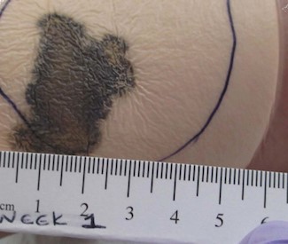
Fig.1. Circle drawn on cover dressing. If strikethrough goes beyond circle, call provider

  • CTPs are different from autologous skin grafts and one should not expect that it be integrated to the wound bed ("take"). CTP will function as a matrix (scaffold) and some may provide growth factors for wound healing.
  • Some CTPs will show a caramelized appearance 5-7 days after application (Figure 2) CTP adhered to wound bed should not be removed

Fig.2. Caramelized appearance 1 week post application of CTP

Videos

Video 1. Wound bed preparation: debridement of non-viable tissue. By S. Robinson, M.D. 

Video 2. Securing the CTP. By S. Robinson, M.D. 

A. Application of Composite Matrix

Case I: Apligraf

Photos below illustrate application and 1 week follow-up of a living, bi-layered CTP made of human keratinocytes and fibroblasts supported by a scaffold (Apligraf). Click on pictures to enlarge.

Fig.A1. Ensure adequate wound bed preparation and hemostasis                                                                                            

Fig.A2. Prepare materials (scissors, forceps, applicator, 11-blade scalpel, silicone sheet, skin closure strips)           
Fig. A3. Apply within 15 minutes from opening the package. Gently lift CTP from medium with forceps or cotton tip applicator. 

Fig.A4. Place CTP on saline-moistened gauze, fibroblast layer (shiny surface)  facing up

Fig.A5. Place gauze with CTP on the CTP dish and fenestrate with a 11 blade scalpel

Fig.A6. Cut CTP to size with scissors

Fig.A7. Hold the gauze with the CTP and apply the CTP on the wound (shiny surface of CTP in contact with wound)

Fig.A8. Peel away gauze while holding CTP in place

Fig.A9. Apply CTP so as to overlap wound edges and ensure wound bed is entirely covered

Fig.A10. Remove air pockets between the CTP and the wound bed. 

Fig.A11. Apply wound glue in the periwound to secure silicone contact layer

Fig.A12. Apply silicone contact layer (e.g, Adaptic, Mepitel) on top of the CTP, covering periwound as well

Fig.A13. Secure contact layer with skin closure strips
Fig.A14. CTP covered with silicone contact layer and secured with skin closure strips. Apply absorptive dressing if exudate

Fig.A15. Follow-up 1 week post CTP application. Secondary absorptive dressing removed. Silicone contact layer visible, on CTP

Fig.A16. Silicone contact layer removed

Fig.A17. Wound with no signs of infection, ready for another application (up to 5 weekly applications)


B. Application of Human Skin Allograft

Case I: AlloPatch Pliable

Photos below illustrate application of human reticular acellular dermal graft (AlloPatch Pliable). Click on pictures to enlarge.

Fig.B1. Prepare  materials (sterile forceps, scalpel, scissors, silicone contact layer)                                                                                               

Fig B2. Debridement of wound bed with curette, removal of fibrin                             

Fig B3. Debridement and hemostasis complete, wound bed ready for CTP application                                                                                                      

Fig.B4. Placement of reticular dermis on wound, cut CTP to size          

Fig.B5. Application of silicone contact layer (e.g, Adaptic, Mepitel) on CTP. Gauze placed between toes to absorb exudate and avoid skin maceration


C. Application of Allogeneic Matrix

Case I : EpiFix

Photos below illustrate application of dehydrated amnion/chorion membrane composed of a single layer of epithelial cells, a basement membrane and an avascular connective tissue matrix (EpiFix). Click on pictures to enlarge.

Fig.C1. Appearance 1 week after application of CTP, preparing wound for CTP reapplication.                                                                                                                                                                                                                    

Fig.C2. Meshed configuration of the CTP is applied on the ulcer so as to overlap wound edges by 1-2mm                                                                                                                                                                                                                     

Fig.C3. Non-adherent silicone contact layer (e.g., Silflex) is applied over the CTP. White color helps post care instructions (only dressing above the "white layer" can be changed before 1 week)                                                                            

Case II: AmnioBand

Photos below illustrate application of an antimicrobial dressing over a dehydrated human placental membrane  (AmnioBand). Click on pictures to enlarge. 

Fig.C4. Non-healing ulcer on paraplegic 20 year-old patient who scooted around the house on ventral decubitus. CTP applied on ulcer*, covered with silicone contact layer and antimicrobial dressing (blue). 
Fig.C5. External foam dressing cover applied over antimicrobial dressing (blue) to provide extra padding and protect from eventual trauma caused by patient's habitual behavior          

* For highly exudative wounds, prior to its application, AmnioBand (in a dry state) can be meshed or fenestrated by small slits through the graft to facilitate drainage. While on the wound site, if needed, hydrate AmnioBand with sterile saline solution. AmnioBand should be covered with a non-adherent dressing as a primary dressing (e.g., Mepitel, Adaptic Touch, Xeroform), followed by a secondary dressing to maintain moisture (e.g. Tegaderm, Kerlix, 3M Coban, etc).

D. Application of Acellular Matrix

Case I: PuraPly Antimicrobial 

Photos below illustrate application of a CTP comprised of purified porcine intestinal collagen type I matrix and the antimicrobial polyhexamethylene biguanide (PHMB) (PuraPly Antimicrobial). Click on pictures to enlarge.

Fig.D1. CTP applied on heavily exudative wound                                                                                                                                   

Fig.D2. CTP is covered and secured with non-adherent silicone contact layer                                                                                         

Fig.D3. Cross-sectional cut illustrating dressing layers. CTP covered with contact layer, covered with superabsorbent dressing (e.g. Eclypse Boot, Mextra)

Case II: Kerecis

The video below illustrates application of fish skin for wound management (Kerecis™ Omega3 Wound)

Video 3. Application of fish skin acellular matrix on a diabetic foot ulcer. By S. Robinson, M.D. 

HOW TO ASSESS EFFECTIVENESS

Upon follow-up, usually 1 week after CTP application, assess: 

  • Signs of improvement, which may be subtle: wound edge epithelialization, decreased wound dimensions (diameter, depth), decreased exudate, inflammation, swelling, pain, necrotic tissue/slough
  • Signs of worsening or infection: absence of these signs may indicate that treatment with CTP is being effective. Signs of infection include [1][2]:
    • NERDSNonhealing, increased Exudate, Red and friable tissue, Debris, Smell. If any 3 = local infection. 
    • STONEES: increased Size, increased Temperature, Os (Orifice), New  breakdown, increased Exudate, increased Erythema/edema (cellulitis), Smell. If any 3 = deep infection
  • Different products may have different appearances 1 week after application. For instance:
    • Living bi-layered CTP (Apligraf) may appear caramelized (Figure A15). This appearance is expected and adhered CTP should not be debrided.
    • CTP comprised of collagen matrix with antimicrobial (e.g., PuraPly Antimicrobial) may be not be visible anymore (i.e., appears "consumed"), indicating presence of high levels of bioburden, warranting further application of this antimicrobial CTP. 
  • Application of CTP should be discontinued when clinician deems that an appropriate response has been obtained or if side effects occur.

TROUBLESHOOTING

Pain

  • Conservative sharp wound debridement (CSWD) performed prior to application of CTP can result in pain. For management of pain due to CSWD, refer to the section 'Troubleshooting' in topic "How to Perform Conservative Sharp Wound Debridement".

Bleeding

  • Bleeding may occur as a result of CSWD performed prior to application of CTP. For management of bleeding due to CSWD, refer to the section 'Troubleshooting' in topic "How to Perform Conservative Sharp Wound Debridement".

Allergic reaction

  • Several CTPs are derived from animal sources. Porcine-derived small intestinal submucosa, porcine urinary bladder matrix, bovine dermis, equine pericardium, and sheep tissue are processed for use as skin substitutes because of their type 1 collagen content.[3] Some patients may have an allergic reaction to animal-sourced products. Use of these products on patients with known sensitivity or allergy is contraindicated.
  • Diagnosis: is often clinical, through history and physical examination. May include local signs such as rash, pruritus and swelling of the periwound area, along with systemic signs. 
  • Management:
    • If an allergic reaction develops on a patient with no previous known sensitivity or allergy, discontinue the product immediately. 
    • Symptoms of allergic contact dermatitis are often managed with topical steroids. Antihistamines may be used to control itch. 
      • For areas of thinner skin such as face, intertriginous areas and periwound, low-potency topical steroids can be used, for a maximum of 14 days (e.g. hydrocortisone 1% or 2.5%, cream or ointment).

Infection

  • Diagnosis: mainly clinical. See signs and symptoms of infection above in section 'How to Assess Effectiveness'.
  • Management:
    • Discontinue CTP.
    • Collect a wound swab. See topic "How to Collect a Wound Swab (Levine Technique) for Culture".
    • Treat local infection (three or more NERDS criteria) with topical antimicrobials (silver, iodine, PHMB/chlorhexidine, methylene blue/crystal violet, surfactants).[4]
    • Consider treating deep and surrounding infection (three or more STONEES criteria) with systemic antimicrobials. May start with empiric antibiotic therapy according to facility protocol, and then implement susceptibility-guided therapies once culture results are available.[4]

REVISION UPDATES

DateComments
1/08/22Version 3.0: added videos, Troubleshooting section, updated protocol and cases


    Official reprint from WoundReference® woundreference.com ©2024 Wound Reference, Inc. All Rights Reserved
    Use of WoundReference is subject to the Subscription and License Agreement. ​
    NOTE: This is a controlled document. This document is not a substitute for proper training, experience, and exercising of professional judgment. While every effort has been made to ensure the accuracy of the contents, neither the authors nor the Wound Reference, Inc. give any guarantee as to the accuracy of the information contained in them nor accept any liability, with respect to loss, damage, injury or expense arising from any such errors or omissions in the contents of the work.

    REFERENCES

    1. Sibbald RG, Woo K, Ayello EA et al. Increased bacterial burden and infection: the story of NERDS and STONES. Advances in skin & wound care. 2006;volume 19(8):447-61; quiz 461-3.
    2. Woo KY, Sibbald RG et al. A cross-sectional validation study of using NERDS and STONEES to assess bacterial burden. Ostomy/wound management. 2009;volume 55(8):40-8.
    3. Agency for Healthcare Research and Quality (AHRQ). Skin Substitutes for Treating Chronic Wounds - Technology Assessment, Technical Brief, Project ID: WNDT0818 . 2020;.
    4. Sibbald RG, Elliott JA, Persaud-Jaimangal R, Goodman L, Armstrong DG, Harley C, Coelho S, Xi N, Evans R, Mayer DO, Zhao X, Heil J, Kotru B, Delmore B, LeBlanc K, Ayello EA, Smart H, Tariq G, Alavi A, Somayaji R et al. Wound Bed Preparation 2021. Advances in skin & wound care. 2021;volume 34(4):183-195.
    Topic 1070 Version 3.0

    RELATED TOPICS

    This topic provides practical guidance to wound care clinicians on the selection of Negative Pressure Wound Therapy (NPWT) devices, device application with step-by-step pictures, assessment of therapy effectiveness, and troubleshooting.

    This topic provides guidance on selection of Cellular and/or Tissue Products (CTPs) with an interactive framework

    t
    -->